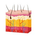
cross section illustration human skin layers the epidermis dermis and |
||||||||||||||||||||||||||||||||||||||||
navigate by keyword : yellow visible vessels varying toy tissues tissue textile sweat subcutaneous structures skin shades sebaceous represent red purposes present play pink pattern outlined organ orange nerve layers layer human hair glands generated furniture function food font follicles fibers features epidermis educational drawing detailed design dermis depicted crosssection components clothing circle cells cartoon brown brand blue blood barrier art adipose |
||||||||||||||||||||||||||||||||||||||||
|
||||||||||||||||||||||||||||||||||||||||
Cross-section illustration of human skin layers. The epidermis, dermis, and subcutaneous tissue are depicted. The epidermis contains cells with a barrier function. The dermis features red and blue blood vessels, nerve fibers, and sweat glands. Yellow adipose tissue is present in the subcutaneous layer. Hair follicles and sebaceous glands are also visible. Varying shades of pink and yellow represent different tissues and components, outlined with detailed structures for educational purposes. |
||||||||||||||||||||||||||||||||||||||||
Stockphotos.ro (c) 2025. All stock photos are provided by Dreamstime and are copyrighted by their respective owners. |